The Capabilities of Ultrasound for Knee Pain Diagnosis
Posted On: November 7, 2025 by Robert Matijevich
Knee pain can be terribly debilitating, and it disrupts your entire lifestyle! You can’t walk comfortably or even perform your usual daily activities. Treatment should begin immediately, but first, a diagnosis is necessary. If the problem is misdiagnosed, you’ll simply waste money on treatment and not get the relief you seek. Moreover, time can be lost. That’s why we recommend a high-quality ultrasound for knee pain diagnosis. Experience the enormous benefits of this innovative diagnostic method.
Prevalence of Knee Pain
Worldwide, the prevalence of knee pain ranges from 10% to 60% across different population groups. The prevalence of knee pain increases with age, reaching 50% in individuals over 75.
In the United States, approximately 25% of adults over 45 experience frequent knee pain.
Anterior knee pain remains one of the most common and complex complaints associated with musculoskeletal disorders. It most often affects individuals between 15 and 30 and is especially prevalent in women. However, men are also susceptible. Despite extensive research, the exact pathophysiology and factors contributing to the development of the disease remain poorly understood. Persistent pain can lead to chronic discomfort, decreased athletic performance, and decreased quality of life.
The etiology of anterior knee pain is multifactorial. Many cases are associated with patellar pathology, particularly patellar instability. However, structural abnormalities alone are not always the cause of pain. Imaging and clinical data often reveal minimal correlation between patellar displacement and long-term treatment outcomes.
Modern Ultrasound Osteoarthritis Assessment
Ultrasound imaging has become one of the primary diagnostic tools for assessing knee joint diseases and traumatic injuries. Ultrasound technologies are constantly being improved, and their accuracy is increasing. Many specialists have already appreciated the high clinical value of this method.
Doctors use this diagnostic method to identify degenerative-dystrophic diseases, inflammatory changes, and vascular and fluid abnormalities in the joint cavity. Specialists also widely use ultrasound to visualize Baker’s cysts, soft tissue neoplasms, and structural damage to the meniscus, tendons, muscles, and ligaments.
This method is completely safe, non-invasive, and has minimal contraindications. Overall, it is a fast, informative diagnostic method. In the presence of contraindications to magnetic resonance imaging, ultrasound remains the optimal alternative.
Modern ultrasound examination of the knee joint is a highly informative and effective method for detecting degenerative and traumatic pathologies. It can be used as a standalone express diagnostic tool or as part of a comprehensive examination, combining it with X-ray, CT scan, or MRI.
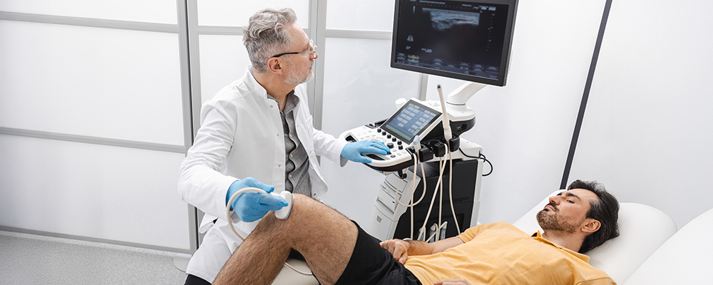
How Does a Knee Ultrasound Work?
It’s a non-invasive, painless, and radiation-free diagnostic method. Modern ultrasound diagnostics offer a fast, accurate, and completely safe way to evaluate a wide range of soft tissue conditions affecting the knee joint. This method uses high-frequency sound waves to produce high-resolution images of the soft tissues surrounding the knee joint.
This technology allows doctors to clearly visualize and evaluate:
- Tendon and ligament tears.
- Meniscus injuries.
- Muscle strains and tears.
- Thickening and inflammation of the synovial membrane.
- Bone erosions associated with rheumatoid arthritis.
- Localized soft tissue lesions or masses.
Results are available almost immediately. The doctor can quickly identify the problem and begin treatment without delay, promoting the best possible recovery outcome.
The Effectiveness of Ultrasound Examination for Knee Pain
Many patients complain of anterior knee pain, which is one of the most common reasons for consulting an orthopedist for knee conditions. Unfortunately, determining its exact etiology remains difficult. Researchers decided to evaluate the diagnostic effectiveness of ultrasound, which was assumed to be a fast and reliable knee imaging technique.
The researchers conducted a prospective analysis involving 143 patients with clinically diagnosed anterior knee pain. Each participant underwent ultrasound and magnetic resonance imaging (MRI) of the affected knee.
A total of 155 knee joints were examined. In 26 cases, specialists found no pathology. The researchers obtained 19 positive MRI results and 110 similar results on ultrasound and MRI. The most common pathology was joint effusion (38%). Patients also suffered from trochlear cartilage defects (20.6%) and superficial infrapatellar edema (20%). Ultrasound demonstrated an overall sensitivity of 85.3% and a specificity of 100%. The highest detection sensitivity was observed for bipartite patella (100%), joint effusion (91.5%), and quadriceps tendinopathy (87.5%).
The hypothesis was confirmed. Ultrasound is a fast, accurate, cost-effective method for assessing anterior knee pain. It can serve as an effective alternative to MRI, especially in cases where access to MRI is limited or contraindicated.
Fast and Accurate Diagnosis of Sports Injuries
Knee injuries are among the most common sports problems faced by professional athletes and active individuals. They can occur due to overuse, sudden twisting, or falls. Common conditions include anterior cruciate ligament (ACL) tears, meniscus injuries, muscle or tendon tears, tendinosis, and overuse syndrome.
Ultrasound imaging allows for quick and accurate diagnosis of these injuries. This helps doctors develop targeted treatment plans that promote healing, reduce rehabilitation time, and restore mobility, allowing you to return to training or competition faster.
Ultrasound-Guided Knee Injections
Ultrasound guidance significantly improves the accuracy and safety of knee injections, ensuring the needle is placed precisely at the desired location. Physicians may supplement certain therapeutic procedures with ultrasound guidance. Autologous blood and platelet-rich plasma injections stimulate the body’s natural healing response. Physicians use sclerotherapy and autologous tenocyte implantation to relieve pain and treat chronic tendinosis. Ultrasound visualizes the injection site in real time. This ensures direct delivery of the medication or regenerative solution to the affected area, minimizes damage to surrounding tissue, and improves overall treatment outcomes.
Key Advantages of Ultrasound in Knee Pain Diagnosis
A comprehensive clinical examination remains the foundation of AKP diagnosis.
MRI has traditionally been the preferred method for assessing the knee joint due to its excellent soft tissue contrast and the ability to visualize multiple joint compartments in detail. It has largely replaced diagnostic arthroscopy for many knee pathologies. However, MRI has limitations: high cost, longer scanning times, and contraindications for some patients (e.g., those with pacemakers or metal implants).
On the other hand, ultrasound offers a noninvasive, fast, and affordable alternative. It provides dynamic, real-time visualization of the soft tissues of the anterior knee, including tendons, bursae, ligaments, and periarticular structures, which can be major sources of pain. Furthermore, ultrasound can be performed at the patient’s bedside, does not expose patients to ionizing radiation, and allows for immediate comparison with clinical data during the examination. MRI remains the gold standard in medicine, but ultrasound allows for rapid assessment of anterior knee pathology. Ultrasound provides high diagnostic accuracy for most structural causes of AKD. Physicians should use MRI if cartilage defects are suspected or if ultrasound results are inconclusive.
Conclusions
Knee joint ultrasound evaluation has become integral to modern sports medicine and orthopedic diagnostics. Doctors now quickly understand the causes of knee pain and injuries. This helps them prescribe targeted treatment and promotes faster and safer recovery. If you’re suffering from knee pain, don’t delay. Get diagnosed and quickly restore joint mobility and relieve knee pain. Mossy Creek Rehabilitation Center provides high-quality physical therapy services in a friendly atmosphere for residents of Jefferson City and the surrounding area. Our staff is committed to helping our patients improve their lives, regain their activity, and feel full again. Contact us for a consultation.